Connall's Heart Description
![]()
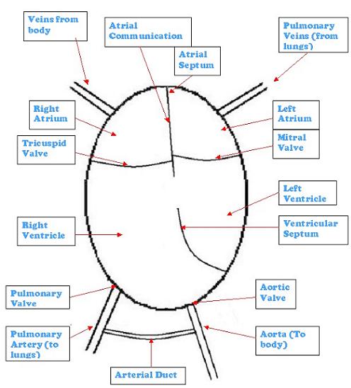
Shown below is Connall's Heart

Straight away there is a big difference between Connall's heart and a normal one.
The first main problem is that the left hand side of the heart (both the left atrium and ventricle) are much smaller than normal. This, along with other problems, is known as Hypoplastic Left Heart Syndrome (HLHS).
With Hypoplastic Left Heart Syndrome, not only are the left atrium and ventricle small, but the mitral, and or aortic valves may be narrow, blocked or not formed properly. The Aorta is often small and not fully formed and there is a hole (Atrial Septum Defect, ADS) between the two atrium's (collecting chambers).
This makes the blood's journey very different from normal. The blood returning from the body, flows into the right atrium (collecting chamber), through the tricuspid valve and into the right ventricle (pumping chamber). From there it is pumped to the lungs, where the blood is oxygenated.
The oxygenated blood then flows back to the left atrium (collecting chamber), from the lungs, but it is unable to pass into the left ventricle (pumping chamber) as the mitral valve will be blocked, it therefore passes through the hole in between the two atrium's, where it mixes with the deoxygenated blood and follows the normal path to the lungs.
Whilst the arterial duct (ductus arteriosus) is still open, the blood will pass from the pulmonary artery into the aorta (body artery) and then around the body. When the duct closes the baby will no longer have oxygen flowing to their body, and gradually they will become sicker and die.
In Connall's case, his problems are compounded by other things that are wrong.
Instead of having a hole in the atrial septum, there (at his 1st scan at Great Ormond Street Hospital or GOSH) was none. This causes the blood to congest when it returns from his lungs in the pulmonary veins, which would cause the structure of the to be muscular and not very flexible. Although 3rd scan at GOSH there did appear to be a small amount of blood flowing through the atrial septum and a decrease in congestion of the pulmonary veins, but also there appeared to be a small collection of fluid on one of this lungs, this might be related, but we are not sure as yet.
Instead of having an intact ventricular septum (wall between the left and right ventricles) there was hole, this is known as a ventricular septum defect or VSD.
His mitral valve (between the left atrium and ventricle) at his 1st scan at GOSH appear to be intact, although again like the atrial septum, at his 2nd and 3rd scans the appear to be a slight improvement which allowed a small amount of blood to flow through.
Another problem that he has is a condition where the pulmonary and aorta arteries are transposed (swapped position). This is known as Transposition of the Great Arteries or TGA. And because his body needs the greater amount of oxygenated blood than his lungs, the pulmonary artery is very small, compared to what it should be in a normal heart.
![]()
![]() What Could Be Done For Connall
What Could Be Done For Connall
![]() Return To Connall's Condition Main Page
Return To Connall's Condition Main Page![]()
![]() Return To Connall's Condition Introduction
Return To Connall's Condition Introduction![]()
![]() Return To Normal Heart Description
Return To Normal Heart Description![]()
29 August, 2004