Normal Heart Description
![]()
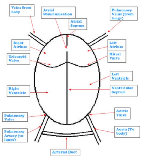
Shown below is the basic diagram of a heart.

The main heart function is to circulate blood through the body and lungs, in two separate circulations (One circuit being the body, the second being the lungs). In a normal heart, as above, the way the blood flows is as follows (taken from the blood flowing into the heart from the veins from the body).
1, The blood flows into the right atrium (collecting chamber) from the body, via the veins that return to the heart.
2, The right atrium then passes the blood through the tricuspid valve into the right ventricle (pumping chamber).
3, The right ventricle then pumps the blood through the pulmonary valve into the pulmonary arteries which go to the lungs.
4, The blood is then oxygenated as it travels through the lungs.
5, The oxygenated blood returns to the heart, via the pulmonary veins into the left atrium (collecting chamber).
6, The left atrium then passes the blood through the mitral valve into the Left Ventricle.
7, The left ventricle then pumps the blood through the aortic valve into the aorta arteries which go to the body. And the cycle begins again.
In babies that are in the womb, obviously there is no air for them to breath in, unlike those on the outside, so the oxygen is passed through the placenta from mummy, instead of through the lungs. Only a small amount of blood is needed to travel through the lungs whilst baby is in the womb. If too much blood passes through the lungs the structure of the lungs and the pulmonary veins and arteries can become muscular. To help avoid this two communication structures are present, which both allow the passing of blood. After birth these close up over the course of a few days or weeks, allowing the baby to develop a proper circulatory system These are a hole in the atrial septum and the arterial duct.
The atrial septum hole or as it is called the atrial communication allows oxygenated blood to flow from the right atrium to the left atrium, where it gets pumped around the body.
The arterial duct or ductus arteriosus as it is also called, joins both the pulmonary arteries (lung arteries) and the Aorta (body arteries). As the blood is pumped up the pulmonary arteries some blood passes the lungs but most flows through the duct to the aorta (body arteries) and back round the body, again avoiding the lungs.
Connall's heart is very different from this,
![]()
![]() Return To Connall's Condition Main Page
Return To Connall's Condition Main Page![]()
![]() Return To Connall's Condition Introduction
Return To Connall's Condition Introduction![]()
30 April, 2005